This site only uses technical cookies necessary for it to work. No analytics or advertising cookies. More info in our Privacy Policy.
We restore the lost volume of the mid-face and cheekbones with your own fat. A younger, more natural face. Permanently.
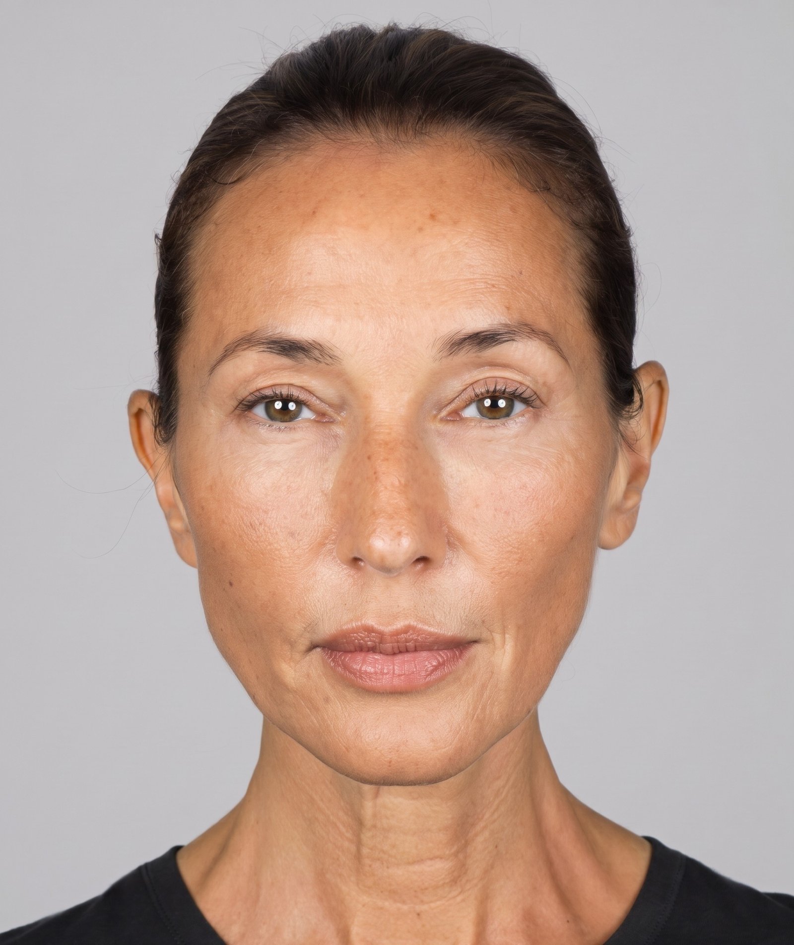
Facial lipofilling extracts fat from the patient's own body and transfers it strategically to the face to restore volume lost with ageing.
The main treatment area is the mid-face and cheekbones. By restoring their projection, features lift and grooves soften naturally, without touching them directly.
This is not superficial filler: it is a structural correction in depth.
No implants or synthetic materials. Zero risk of rejection or allergic reaction.
Integrated fat lasts a lifetime. No periodic maintenance.
The donor area improves and the face rejuvenates in a single procedure.
Stem cells stimulate collagen, improving texture and radiance.
By integrating as own tissue, the result is organic and harmonious.
Can be combined with blepharoplasty or aesthetic medicine for an integral result.
"Dr. Jiménez is a true professional and his team is exceptional. Even before the swelling went down, the change was visible."
"He perfectly understood my needs. He exceeded my expectations. The professionalism of the whole team is outstanding."
"I had a complete blepharoplasty and it was a complete success. I am very satisfied and highly recommend it."
"I had surgery in December and I couldn't be happier. From day one you notice the results. I would choose him again."
We analyse your case personally and without commitment.
What it consists of, which areas it treats, how it is performed and what to expect.
Facial lipofilling extracts fat from the patient by gentle liposuction, purifies it in the operating room following the Coleman protocol and reinjects it strategically into the face.
The result is three-dimensional rejuvenation with a lifting effect across the entire mid and upper face: the gaze is lifted, lost volume is recovered and skin quality improves thanks to the growth factors and stem cells in fat tissue.
Local with mild sedation. Outpatient: you go home the same day.
Between 1 and 2 hours depending on areas and volume needed.
Presentable at 7 days when no surgery is combined: cannula only, no incisions, no stitches.
From 3–6 months. Integrated fat is permanent.
Restoring their projection lifts the entire face, narrows dark circles and reduces nasolabial folds without touching them directly.
Fills the tear trough from depth, with more natural results than superficial fillers.
Temporal hollowing is a frequent and under-diagnosed sign of ageing.
Grafting at the brow tail and temporal region lifts and supports the upper third: the gaze opens up without traction surgery.
Filling of the hollowed upper eyelid and deep orbital sulcus, and correction of a sunken eye: post-traumatic, after orbital surgery or in ocular prosthesis wearers.
Emulsified fat (nanofat), rich in regenerative cells, is injected superficially to improve texture, radiance and fine lines, and to treat acne or surgical scars.
Projection and definition of the lower facial profile.
Complete morphological analysis and personalised design. First consultation always free.
Minimally invasive liposuction from the donor area under local anaesthesia.
Gentle decanting — no centrifugation — to avoid adipocyte rupture and maximise cell survival.
Microdroplet by microdroplet injection in multiple planes with blunt microcannulas.
Reviews with Dr. Jiménez Ortiz. 24h emergency telephone assistance.
A periocular surgery technique aimed at restoring lost volume around the eyes — upper eyelid, lower eyelid and the orbitomalar groove — using the patient's own fatty tissue, performed by an oculoplastic surgeon.
Ageing around the eyes is not a single phenomenon: it produces skin changes, muscle changes, changes in the supporting ligaments, orbital bone remodelling and redistribution (with loss) of periocular fat, all at once. A purely resective blepharoplasty — which only removes skin and fat — can be insufficient, or even counterproductive, in patients where volume loss is the main component.
A small amount of autologous fat is harvested from a donor area through gentle liposuction.
The fat is gently decanted (without centrifuging) to preserve the adipocytes and the cellular fraction.
It is infiltrated in micro-deposits, into selected areas of the periocular contour, using a microcannula.
Fat grafting does not work like a simple filler. The transferred fat behaves like a living tissue graft: once implanted, it has no vascularisation of its own and needs to integrate with the recipient tissue to survive.
Rather than concentrating large amounts of fat at a single point, the graft is distributed into small deposits, across different planes and pathways when anatomy allows it. This increases the contact surface between the fatty tissue and the vascularised recipient tissue, which is what determines initial survival.
The reference histological model (Eto et al., 2012) describes three zones within a fat deposit: a peripheral survival zone (in direct contact with recipient tissue), an intermediate regenerative zone (where adipocytes die but progenitor cells survive and regenerate new tissue) and a central necrotic zone when the deposit is too large to receive oxygen by diffusion.
Simplified conceptual diagram of the integration process of a fat micro-graft. It does not represent an exact time scale.
Part of the transferred volume may be reabsorbed during the first few months. The final volume depends on several factors, not only on the surgical technique:
Swelling and possible bruising. The visible volume is greater than what will remain permanently.
The apparent volume progressively decreases. It still does not equal the final result.
Main phase of neovascularisation and remodelling of the graft.
The result is now much more reliable, although it may continue to refine.
Fine biological remodelling may continue for up to approximately 12 months.
Times are approximate and vary depending on the procedure and each patient.
Indicated for a deep superior sulcus, a skeletonised eyelid appearance, age-related loss of fullness, sequelae of previous overly resective blepharoplasties, or volume asymmetries between both eyes.
Tear troughs can have very different causes: skin pigmentation, thin skin, a herniated fat pocket, bony or soft-tissue hollowing, or a combination of several. Fat grafting only makes sense when there is a real component of volume loss.
Fat grafting and blepharoplasty are not mutually exclusive: they can be combined in the same surgical procedure. Modern periocular surgery is not only about resecting; in selected patients it also involves repositioning tissue and restoring volume.
It preserves structural adipose tissue: it retains adipocytes and provides real volume. Its main goal is three-dimensional restoration. Potential periocular applications: a skeletonised upper eyelid, the orbitomalar groove, the lid-cheek transition, the temporal region and certain sequelae of previous surgery.
Adipose tissue subjected to greater mechanical processing, which destroys most of the mature adipocytes. Its volumising capacity is minimal; the interest is mainly stromal and cellular, under research for tissue quality and scars. Clinical evidence on its regenerative effects is promising but still heterogeneous.
| Microfat | Nanofat | |
|---|---|---|
| Goal | Volume | Tissue quality |
| Adipocytes | Preserved | Mostly altered |
| Volume effect | Yes | Minimal |
| Main use | Restoration | Tissue remodelling |
An upper eyelid hollowed by excessive previous fat resection, irregularities, asymmetries, hollowing or postoperative skeletonisation are frequent reasons for secondary consultation. Revisional cases require an especially careful evaluation: the anatomy may be altered by previous surgery, fibrosis and scarring, which makes the result less predictable.
Fat that successfully integrates can behave like the patient's own adipose tissue, and be potentially long-lasting. It can, however, change over time due to age, weight changes, metabolism or later anatomical changes — just like the rest of the body's fatty tissue.
The choice depends on the anatomy, the indication, the history and the goals of each patient. Neither option is universally superior.
Oedema, bruising, asymmetry, contour irregularity, overcorrection or undercorrection, greater-than-expected resorption, fatty nodules or cysts, and the possible need for a touch-up.
As with other facial injection procedures, the medical literature describes very infrequent but potentially serious vascular complications (including vision loss) associated with the injection of fat or other materials in the periocular and facial region.
The safety margin depends directly on: detailed anatomical knowledge of the periocular region, careful selection of the injection plane, appropriate patient selection, and specific experience in periocular surgery — not only in general aesthetic medicine.
It is obtained through gentle liposuction of a donor area on the patient's own body (usually the abdomen or flanks), under local anaesthesia. The amount needed for the periocular region is small.
There is no fixed figure: in facial fat studies, the published average retention is around 47%, with a range of 26–83% depending on technique and measurement method. It depends on the recipient's vascularisation, the amount transferred and each patient's biology.
It is uncommon, but individual variability exists. Part of the volume is reabsorbed as expected during the first few months; in selected patients a second session may be necessary.
The initial result is not final due to inflammation. It becomes much more representative from 3–6 months onward, and fine biological remodelling may continue for up to approximately 12 months.
Yes, this is a frequent combination in modern periocular surgery. The specific strategy (which area to operate on, which area to fill) is defined after assessing each case.
Neither option is universally superior. Hyaluronic acid is reversible and gives a more immediate result; fat grafting uses the patient's own tissue and is potentially longer-lasting. The choice depends on each patient's anatomy and goal.
No. It only makes sense when there is a real component of volume loss. Tear troughs caused by pigmentation, thin skin or herniated fat pockets may require other treatments, or a combination of them.
Microfat preserves adipocytes and provides real volume; it is used for structural restoration. Nanofat undergoes greater mechanical processing, provides very little volume, and its interest is mainly cellular/stromal, with clinical evidence still heterogeneous.
Yes, in certain patients, especially if resorption was greater than expected. This is not considered a failure of the technique, but a possibility anticipated within the process.
Fat that integrates can behave like the patient's own tissue and be potentially long-lasting, but it can change over time due to age, weight or metabolism — just like the rest of the body's fat.
In certain cases of a skeletonised or hollowed eyelid after previous surgery, fat grafting can improve appearance. Since this is revisional surgery, the altered anatomy makes the result less predictable, and it is assessed individually.
The most frequent are oedema, bruising, asymmetry or irregularity, generally mild and transient. The medical literature also describes serious vascular complications, extraordinarily rare, associated with facial injection. Experience in periocular surgery reduces this risk.
The content of this page is based on published scientific literature on autologous fat transfer. These are some of the most relevant references:
Eto H, Kato H, Suga H, et al. "The Fate of Adipocytes after Nonvascularized Fat Grafting." Plast Reconstr Surg. 2012;129(5):1081–1092. PubMed 22261562
Tonnard P, Verpaele A, Peeters G, et al. "Nanofat Grafting: Basic Research and Clinical Applications." Plast Reconstr Surg. 2013;132(4):1017–1026.
Shih L, et al. "The Science of Fat Grafting." Semin Plast Surg. 2020;34(1):5–10.
Revisión sistemática y metaanálisis. "Volume Retention After Facial Fat Grafting and Relevant Factors." Aesthetic Plast Surg. 2021;45(2):506–520. PubMed 31940073
Yang F, et al. "Efficacy, Safety and Complications of Autologous Fat Grafting to the Eyelids and Periorbital Area: A Systematic Review and Meta-Analysis." PLoS One. 2021;16(4):e0248505. PMC8016360
Khouri RK, Rigotti G, Cardoso E, et al. "Megavolume Autologous Fat Transfer: Part I. Theory and Principles." Plast Reconstr Surg. 2014;133(3):550–557.
Lazzeri D, Agostini T, Figus M, et al. "Blindness Following Cosmetic Injections of the Face." Plast Reconstr Surg. 2012;129(4):995–1012. PubMed 22456369
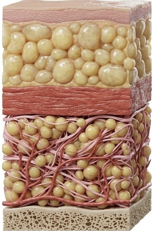
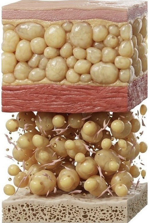
Compare the freshly placed fat graft with the same graft once integrated, when the extracellular matrix has completed angiogenesis.
In the first days the graft survives by diffusion from the recipient bed: it does not yet have its own blood supply.
The recipient tissue generates new vessels that penetrate the graft along the matrix scaffold.
By around 3 months the extracellular matrix has reorganised and the remaining volume stabilises.
"I have bags" is one of the most common reasons for consultation, but it covers very different causes. The right treatment depends on the real cause — not all bags are treated the same way, and not all of them require surgery.
This is the classic cause: with age, the septum that holds periorbital fat in place weakens and the fat protrudes forward, forming a visible bulge under the lower eyelid. It is usually corrected with lower blepharoplasty (removing or repositioning the fat), not with fat grafting.
When the cheekbone or the orbitomalar groove lose volume, the bag —even without having grown— becomes more visible by contrast. This is where fat grafting can help, not by filling the bag itself, but by softening the transition around it.
The skin of the lower eyelid is among the thinnest in the body. With age it can lose elasticity and form folds that are mistaken for fat bags. The approach here is different: skin quality treatment, or skin resection if there is a relevant excess.
A dark tone under the eye is not a volume problem: it can be due to thin skin revealing the underlying vessels, pigment deposits, or the shadow cast by a groove. Fat grafting does not correct pigmentation itself.
Morning puffiness from fluid retention —common due to genetics, sleep, or salt intake— is not a structural problem. It usually improves throughout the day and does not require surgical treatment.
"My eyelids are drooping" describes, in consultation, two anatomically distinct situations — which are treated differently. Telling them apart is the first step of any assessment.
This is the most common: the skin of the upper eyelid loses firmness with age and falls over the crease, sometimes interfering with peripheral vision. The eyelid itself functions normally — the problem is the excess skin. It is corrected with upper blepharoplasty.
This is different: the eyelid margin covers more of the eye than normal because the levator muscle —which holds the eyelid up— is not working with its usual strength, not because of excess skin. Oculoplastic surgeons differentiate it from dermatochalasis through a specific eyelid examination, not just a visual one. Correcting it requires a different technique from standard blepharoplasty.
Both problems can coexist in the same patient, and periocular volume loss —treatable with fat grafting— can visually accentuate the drooping appearance without being its cause. That's why the assessment must consider skin, muscle and volume separately before deciding what to correct.
"Blepharoplasty" doesn't describe a single technique. The right approach depends on whether the main problem is excess skin, herniated fat, or volume loss — and in practice they can be combined.
Removes the excess skin of the upper eyelid —and, if necessary, repositions or removes localised fat— to open up the eyes and, in cases with functional impact, improve peripheral visual field.
Addresses herniated fat and/or excess skin of the lower eyelid. Depending on the case, the fat can be repositioned (rather than removed) to fill the orbitomalar groove from within, avoiding a hollow appearance after surgery.
When the main problem is not excess skin or fat but volume loss —a skeletonised eyelid, a hollow temple, a marked lid-cheek transition—, a non-resective approach can be chosen: restoring volume with fat grafting instead of removing tissue. This approach is sometimes referred to as volumetric blepharoplasty, and is the central focus of this practice in cases where it is indicated.
Blepharoplasty and fat grafting are not mutually exclusive and don't compete with each other: they solve different problems —skin and herniated fat versus volume loss— and in selected patients are combined in the same surgical procedure.
Specialist in ophthalmology and oculoplastic surgery. MIR residency at La Princesa University Hospital, advanced training at Stanford Hospital (Byers Eye Institute, California), in Beverly Hills with Dr. Jalalabadi and at La Paz University Hospital in Madrid.
Over the last 7 years he has developed his own fat grafting protocol that controls every detail — harvesting, decanting and filtering —, adapting the size of the fat graft to each area of the face. The protocol never varies: predictable, consistent, long-lasting results. More than 3,000 rejuvenation procedures performed.
The first consultation is free and without commitment.
Not all profiles are the same. Find out if you are a good candidate.
Lipofilling achieves the best results in people with facial volume loss due to ageing who seek a natural, lasting solution without synthetic materials.
These are not exclusive techniques. Each has its own indications. In many cases the best solution is to combine them.
They are not rivals: they treat different problems. Fat grafting restores lost volume and lifts the face with a lifting effect; a surgical facelift corrects laxity and excess skin.
The procedure is performed under anaesthesia. The post-operative period is manageable with standard painkillers, mild during the first 2–3 days.
Generally between 50 and 100 cc. Most patients, even the slimmest, have adequate reserves. Evaluated during consultation.
At 7 days if fat grafting is not combined with surgery: it is performed with a cannula only, with no incisions or stitches. If combined with blepharoplasty or other surgery, recovery follows that procedure (10–14 days).
Yes, although initial swelling distorts the real volume. The definitive result is appreciated from 3–6 months.
Yes, it is a very common and complementary combination. Dr. Jiménez Ortiz will evaluate the best option.
It is not a surgical facelift (which tightens the skin), but it produces a lifting effect: by restoring volume in the cheeks, temples and midface, the face is lifted and the gaze opens naturally, with no scars. When the issue is volume loss, fat grafting is usually the best option; with significant skin laxity, a facelift or a combination is considered.
It depends on the cause. If it's herniated fat, it's usually corrected with lower blepharoplasty. If the problem is surrounding volume loss, fat grafting can help. If it's skin or pigmentation, the approach is different. That's why diagnosis is the first step, not treatment.
Not directly. Fat grafting does not remove herniated fat — that requires blepharoplasty. What fat grafting can do is fill the area around the bag to soften the transition and improve the overall appearance, when volume loss is also present.
Excess skin (dermatochalasis) is a problem of the eyelid skin, which sags with age; the eyelid itself opens normally. Eyelid ptosis is a problem of the muscle that holds the eyelid up, causing it to cover more of the eye than normal, with or without excess skin. They require assessment and, often, different surgical techniques.
This isn't something you can determine just by looking in the mirror: it requires a specific eyelid examination. During consultation, the eyelid's position relative to the pupil and the levator muscle's function are assessed, along with excess skin, to differentiate both conditions — which often coexist.
It is the surgery that corrects excess skin and/or herniated fat in the upper and lower eyelids. It can be performed on one eyelid, on both, or combined with fat grafting when volume loss is also present.
It depends on where the problem is: excess skin above, herniated fat or skin below, or both. This is determined through an examination in consultation; not all patients need both eyelids operated on.
It is an eyelid rejuvenation approach based on restoring volume with the patient's own fat (fat grafting), instead of removing skin or fat as in classic blepharoplasty. It makes sense in patients where the main component is volume loss, not tissue excess; the right approach for each case is assessed in consultation.
Fat grafting in the tail of the brow and the temple can produce a visual lift of the gaze by restoring lost volume in that area, without surgery or scars. It is not a surgical brow lift (which physically repositions tissue through incisions) and does not replace it in cases of marked sagging — it is a different option, useful when the main problem is volume loss.
Not necessarily. The processing of the fat graft is adapted to the area being treated: on the cheekbones, temples or cheeks, it may be preferable to preserve a larger graft (macrofat) to maximise projection; on the eyelids and other very thin-skinned areas, a finer processing (microfat) is used, with a maximum of 10 passes through the filter to preserve the matrix surrounding the adipocyte, to achieve a natural result without the graft being noticeable. The decision is made individually during the assessment.
How long does it last? What percentage of fat survives? Honest answers.
The great advantage over hyaluronic acid is that the fat that manages to integrate lasts a lifetime, ageing naturally with the patient.
The process requires the injected fat to vascularise. Not all of it succeeds, and the technique is designed to maximise this percentage.
Between 50–70% of grafted fat integrates permanently.
Yes. Coleman technique, multi-plane injections and not smoking maximise the result.
Expected swelling and bruising. Visible volume is greater than the final result.
Without associated surgery the result is already presentable: cannula only, no incisions or stitches. Most people resume their social life.
Volume decreases as non-integrated fat is reabsorbed. Normal and expected.
Integrated fat stabilises. Definitive result visible. Skin improvement evident.
Integrated fat lasts a lifetime. No periodic maintenance.
Compare both images to see why the result takes months to stabilise: the graft needs to build its own blood supply before it can be considered integrated.
In some cases a second session may be recommended, especially if the reabsorption rate was higher than expected.
We always wait for the result to stabilise —minimum 6 months— before considering a second procedure.
Of the grafted fat integrates permanently. The percentage varies by area, technique and patient habits.
Programs taught by Dr. Antonio Jiménez Ortiz in Madrid: operating-room observership courses and hands-on courses with real patients.
The Instituto de Cirugía Oculofacial offers practical facial fat grafting training for physicians who want to add this technique to their practice. All teaching takes place during real surgery, in an accredited operating room, in small groups.
The program covers the full process following the Coleman protocol: patient selection, treatment planning, harvesting, processing and zone-by-zone injection, plus complication management and follow-up.
Small groups of 2–4 physicians per edition.
Madrid · The Institute's accredited operating room.
Spanish and English.
Held periodically. Ask about the next available dates.
Looking to refine facial fat transfer and its aesthetic planning.
Interested in tear-trough and periocular volume restoration.
Wishing to expand their portfolio with minimally invasive surgical facial rejuvenation.
Especially those focused on rhinoplasty and facial surgery who want to add autologous fat grafting.
Skilled injectors ready to move to permanent volume with autologous fat.
Watch live facial fat grafting surgeries performed by Dr. Jiménez Ortiz, with step-by-step explanation of every technical decision.
Perform the technique end to end on a real patient, under the direct supervision of Dr. Jiménez Ortiz, on cases pre-selected by the Institute.
Fat compartments, danger zones and prevention of vascular complications.
Morphological analysis, photography, indications and limits of the technique.
Donor sites, tumescent infiltration and atraumatic liposuction.
Atraumatic decanting — no centrifugation, to avoid adipocyte rupture — graft preparation and obtaining microfat and nanofat.
Cheek, lid–cheek junction, temple, chin and jawline. Planes, cannulas and volumes.
Prevention and management of complications. Case discussion and postoperative protocol.
Specialist in ophthalmology and oculoplastic surgery. MIR residency at La Princesa University Hospital, with training at Stanford (Byers Eye Institute), Beverly Hills (Dr. Jalalabadi) and La Paz University Hospital. More than 3,000 rejuvenation procedures performed.
Creator of his own fat grafting protocol, refined over 7 years and never varied, to deliver consistent results. Director of the Instituto de Cirugía Oculofacial in Madrid, teaching associate at the Autonomous University and international speaker at congresses and courses.
Training built on a proprietary protocol refined over 7 years and on the published evidence on facial fat transfer.
First consultation always free and without commitment. We attend you personally.